News Story
New Imaging Technique Covers Middle Ground Between Microscopy and Macroscopy
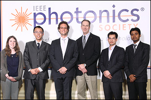
The winners of the IEEE Photonics' Student Paper Awards. Chao-Wei Chen (ECE), advised by BioE assistant professor Yu Chen, is second from the left. IEEE President James Coleman is third from the right.
Department of Electrical and Computer Engineering graduate student Chao-Wei Chen, advised by Fischell Department of Bioengineering (BioE) professor Yu Chen, presented the work, which was conducted in collaboration with BioE professor John Fisher and his advisee, BioE graduate student Andrew Yeatts.
"The research is about filling the gap between microscopy and macroscopy," says Chao-Wei Chen. "Macroscopy techniques such as MRI or CT provide great penetration, greater than 50cm, but are not able to provide great resolution, only about 0.5mm. Microscopy, other the other hand, provides excellent resolution to less than 0.5 microns, but fairly shallow penetration into biological samples, only up to 500 microns, especially working in fluorescence mode."
The system the team created, which has been named "angled fluorescence laminar optical tomography" (aFLOT), offers a balance of penetration and resolution designed to provide quality images of biological structures whose sizes and locations fall somewhere between microscopy's and macroscopy's ideal conditions—in this case, stem cells measuring about 50 microns.
With a 50-100 micron resolution and 2-3mm penetration depth, the device enabled the team to non-invasively obtain three-dimensional images of the distribution of fluorescent-labeled, live stem cells growing in engineered tissue. Currently, a tissue sample would need to be extracted, frozen, sliced into fine sections, scanned and digitally recompiled in order to generate a comparable image. That technique, while effective, is time-consuming and destructive.
Chen and his colleagues consider aFLOT a promising new imaging technique with potential application in regenerative medicine.
The aFLOT project was supported by the National Institutes of Health and the National Science Foundation.
Published January 19, 2012
Recent Stories
Stories / Apr 24, 2024
In Memoriam: Professor Emeritus Moon-Jhong Rhee
Stories / Apr 22, 2024
Khaligh Honored With Linda Clement Outstanding Advisor Award
Stories / Apr 19, 2024
Celebrating Asian, Pacific Islander, and Desi American Engineers

Stories / Apr 16, 2024
Cunxi Yu Receives NSF Medium Award

Stories / Mar 22, 2024
Dinesh Manocha Inducted into IEEE VGTC Virtual Reality Academy
Stories / Mar 21, 2024
ECE Ph.D. Student Ayooluwa (“Ayo”) Ajiboye Recognized at...
Stories / Mar 15, 2024
Fischell Institute Womxn’s History Month Spotlight: Sydney...
Stories / Mar 15, 2024
Agents of Positive Change: Highlighting Women Maryland Engineers

Stories / Mar 14, 2024
Clark School Research Nominated for “Invention of the Year”
Stories / Mar 13, 2024
Edo Waks Recipient of 2024 MURI Award